Cervical Spinal Stenosis
Narrowing of the cervical spinal canal that may place pressure on the spinal cord and nerves
The cervical spine, or neck, plays an important role in supporting the head, protecting the spinal cord, and allowing movement. Cervical spinal stenosis occurs when the space within the spinal canal becomes narrowed, which may place pressure on the spinal cord and surrounding nerves. This narrowing is most commonly related to age-related changes in the spine, including disc degeneration, thickening of ligaments, and the formation of bone spurs. In some cases, it may also be associated with congenital (present from birth) differences in spinal canal size or previous injury.
Cervical spinal stenosis can affect individuals in different ways. Some people may have narrowing visible on imaging without significant symptoms, while others may experience neck pain, arm symptoms, or signs of spinal cord involvement that can affect balance, coordination, and hand function.
At Melbourne Orthopaedic Clinic, cervical spinal stenosis is assessed through a detailed clinical evaluation and appropriate imaging. This allows the underlying cause and severity of narrowing to be understood, helping to guide treatment options tailored to your individual needs.
What is cervical spinal stenosis?
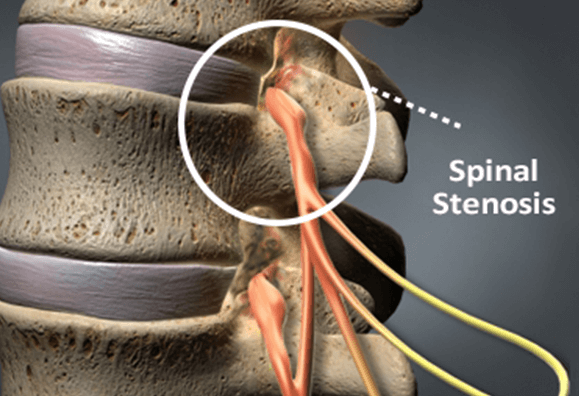
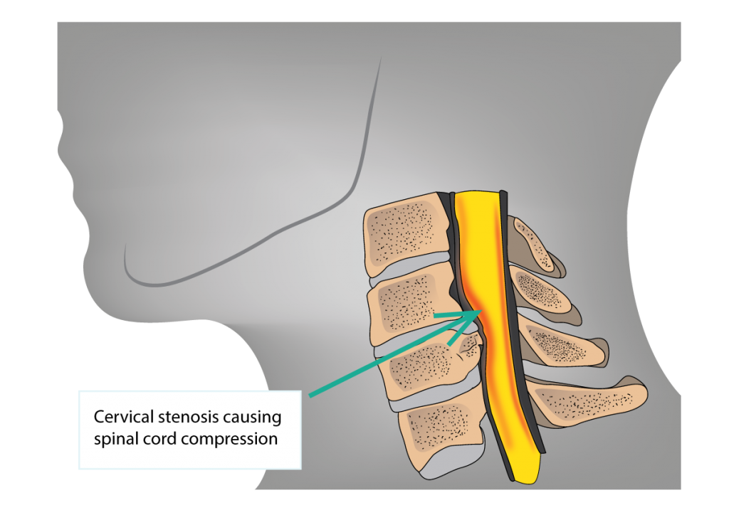
Cervical spinal stenosis refers to a narrowing of the spinal canal in the neck. The spinal canal is the space within the vertebrae that houses and protects the spinal cord. When this space becomes reduced, it may place pressure on the spinal cord and nearby nerve structures.
This narrowing often develops gradually over time as part of the natural ageing process. Changes such as disc degeneration, thickening of surrounding ligaments, and the formation of bone spurs can all contribute to a reduction in space within the spinal canal.
In some individuals, the spinal canal may be naturally narrower from birth, which can increase the likelihood of symptoms developing later in life. Previous injury or other structural changes in the spine may also play a role.
Cervical spinal stenosis can affect people differently. Some individuals may have narrowing seen on imaging without noticeable symptoms, while others may experience neck pain, arm symptoms, or signs of spinal cord involvement. When the spinal cord is affected, this is sometimes referred to as cervical myelopathy, which can influence balance, coordination, and hand function.
Understanding the degree of narrowing and how it relates to your symptoms is an important part of guiding appropriate management.
Common causes and symptoms of cervical spinal stenosis
Cervical spinal stenosis most commonly develops due to age-related changes in the spine, although several factors may contribute to the narrowing of the spinal canal. In many cases, it is a combination of these changes rather than a single cause.
Common causes may include:
- Disc degeneration
Over time, the intervertebral discs can lose height and hydration. This may reduce the space within the spinal canal and contribute to increased pressure on the spinal cord or nerves. - Bone spurs (osteophytes)
As the spine undergoes wear and tear, small bony growths may develop. These bone spurs can project into the spinal canal and contribute to narrowing. - Thickening of ligaments
Ligaments that support the spine, particularly the ligamentum flavum, can become thicker and less flexible over time, reducing the available space for the spinal cord. - Congenital (developmental) narrowing
Some individuals are born with a naturally narrower spinal canal, which may increase the likelihood of developing symptoms later in life. - Previous injury or trauma
Injuries to the cervical spine may alter its structure or stability, contributing to narrowing or inflammation around the spinal cord.
Symptoms of cervical spinal stenosis
Symptoms can vary depending on the degree of narrowing and whether the spinal cord, nerve roots, or both are affected. Some people may have minimal symptoms, while others may experience more noticeable functional changes.
- Neck pain and stiffness
You may experience discomfort or reduced movement in the neck, which can be persistent or intermittent. - Arm pain, numbness, or tingling
If nearby nerves are affected, symptoms may extend into the shoulder, arm, or hand, similar to cervical radiculopathy. - Weakness in the arms or hands
Compression of the spinal cord or nerves may affect muscle strength, making tasks such as gripping or lifting more difficult. - Changes in balance and coordination
When the spinal cord is involved (cervical myelopathy), you may notice unsteadiness when walking or difficulty with coordination. - Reduced fine motor control
Tasks requiring hand precision, such as buttoning clothing or writing, may become more challenging.
Symptoms affecting both sides of the body
Unlike nerve root compression alone, spinal cord involvement may lead to symptoms in both arms or legs.
Symptoms often develop gradually, although in some cases they may progress over time. If you notice changes in strength, coordination, or balance, further assessment may be recommended to better understand the cause and guide appropriate management.
How cervical spinal stenosis is diagnosed
Diagnosing cervical spinal stenosis involves understanding your symptoms, assessing how your spinal cord and nerves are functioning, and identifying the underlying cause and extent of narrowing within the spinal canal.
Assessment typically begins with a detailed clinical history. This includes discussing your symptoms, how long they have been present, whether they are changing over time, and how they are affecting your daily activities, balance, and coordination.
A focused physical examination is then performed to assess neck movement, muscle strength, sensation, reflexes, and coordination. Particular attention is given to signs that may indicate spinal cord involvement (cervical myelopathy), such as changes in balance, hand function, or walking pattern.
Imaging plays an important role in confirming the diagnosis and understanding the degree of narrowing. Investigations may include:
- X-rays: X-rays can provide information about the alignment of the cervical spine and may show signs of disc height loss or bone spur formation.
- MRI (Magnetic Resonance Imaging): MRI is the most commonly used imaging modality for cervical spinal stenosis. It provides detailed views of the spinal cord, discs, and surrounding soft tissues, helping to identify areas of compression or narrowing.
- CT scan (Computed Tomography): A CT scan may be recommended in some cases to provide more detailed assessment of the bony structures of the spine, particularly where bone spurs are contributing to stenosis.
- Additional tests (if required): In selected cases, further investigations may be used to assess nerve function or to differentiate cervical spinal stenosis from other conditions with similar symptoms.
Not all patients will require every test. The choice of investigations will depend on your symptoms, clinical findings, and how the condition is progressing. A clear understanding of both your clinical presentation and imaging findings helps guide appropriate treatment options tailored to your individual needs.
Treatment options and recovery for cervical spinal stenosis
Management of cervical spinal stenosis is tailored to your individual symptoms, the degree of spinal canal narrowing, and whether there is involvement of the spinal cord or nerve roots. In many cases, treatment begins with non-surgical approaches, with surgery considered where symptoms are more significant or progressive.
Non-surgical treatment options
For mild to moderate symptoms, non-surgical management may help reduce discomfort and support function:
- Activity modification: Adjusting positions or activities that aggravate symptoms may help reduce strain on the cervical spine.
- Pain management strategies: Simple pain relief or anti-inflammatory medications may be recommended under the guidance of your GP or treating practitioner.
- Physiotherapy: A structured physiotherapy program may help improve neck mobility, strengthen supporting muscles, and support posture. This can assist in reducing pressure on affected structures.
- Exercise and rehabilitation: Targeted exercises may help maintain movement and function. These are usually progressed gradually based on your response.
- Image-guided injections (in selected cases): In some situations, injections may be considered to help reduce inflammation and provide temporary symptom relief.
Surgical treatment options
Surgery may be considered if symptoms persist despite appropriate non-surgical care, or if there are signs of spinal cord compression, progressive weakness, or changes in balance and coordination.
The aim of surgery is to relieve pressure on the spinal cord and nerves and, where required, stabilise the spine. The most appropriate procedure depends on the location and severity of stenosis.
Common procedures may include:
- Anterior cervical discectomy and fusion (ACDF): Removal of the affected disc and decompression of the spinal cord or nerves from the front of the neck, followed by stabilisation of the spine.
- Cervical laminectomy or laminoplasty: Procedures performed from the back of the neck to create more space for the spinal cord by removing or reshaping parts of the vertebrae.
- Cervical disc replacement (in selected cases): Replacement of a damaged disc with an artificial implant to maintain motion at that level while relieving compression.
All treatment options, including potential risks and benefits, will be discussed with you to support informed decision-making.
Recovery and outlook
Recovery following treatment varies depending on the severity of the condition and the type of management undertaken. With non-surgical care, symptoms may improve gradually over time, although ongoing monitoring may be recommended to assess for any progression.
Following surgery, recovery typically involves a period of activity modification, followed by a structured rehabilitation program, often including physiotherapy. The focus is on restoring movement, strength, and function while protecting the spine during healing. The degree of improvement can vary between individuals. In cases where the spinal cord has been affected, treatment aims to prevent further progression and may also lead to improvement in symptoms, although this can depend on how long the compression has been present.
Ongoing follow-up is an important part of care to ensure recovery is progressing as expected and to support your return to daily activities.